An echocardiogram, often called a heart ultrasound, is a safe, non invasive test that provides detailed images of your heart. It allows doctors to see how well your heart is pumping, check the structure of heart chambers, and evaluate the function of heart valves.
This test can detect a wide range of heart problems, including heart failure, valve diseases, congenital defects, and blood flow issues.
Many people wonder exactly what an echocardiogram shows and how the results are interpreted.
In this article, we will explore everything you need to know about echocardiograms, including the types of tests available, what conditions they can detect, how to prepare for the procedure, and how doctors interpret the results.
By understanding this test, you can better monitor and protect your heart health.
What is an Echocardiogram?
An echocardiogram is a medical imaging test that uses ultrasound waves to create moving pictures of the heart. The test allows doctors to examine the heart’s structure and function in real-time, providing essential information about how well the heart is working.
During an echocardiogram, a small device called a transducer sends sound waves into the chest. These waves bounce off the heart structures and return as echoes, which are converted into detailed images on a monitor. This allows doctors to visualize the size and thickness of heart chambers, the motion of the heart walls, and the function of the heart valves. Advanced techniques, such as Doppler echocardiography, can also measure the direction and speed of blood flow, helping identify conditions like valve disorders or blood clots.
Echocardiograms are commonly used to diagnose and monitor various heart problems, including heart failure, valve diseases, cardiomyopathy, and congenital defects. They can also be part of routine heart health checks, especially for individuals at higher risk of cardiovascular issues. Because the test is non-invasive, relatively quick, and painless, it is often the first choice for assessing heart function and guiding treatment plans.
Types of Echocardiograms
There are several types of echocardiograms, each designed to provide specific information about the heart’s structure and function. The most common type is the transthoracic echocardiogram (TTE). In this non-invasive procedure, a transducer is placed on the chest to send and receive ultrasound waves. TTE is widely used because it is painless, quick, and provides clear images of the heart chambers, valves, and overall heart function.
Another type is the transesophageal echocardiogram (TEE). For this test, a small probe is gently inserted into the esophagus, which lies close to the heart. TEE produces highly detailed images, making it especially useful for evaluating heart valves, detecting blood clots, or assessing certain congenital heart defects. Patients may need mild sedation during this procedure to ensure comfort.
Stress echocardiograms are used to observe how the heart performs under physical or pharmacological stress. Patients may exercise on a treadmill or receive medication to increase heart activity while images are taken. This type is helpful for diagnosing coronary artery disease and evaluating heart function during activity.
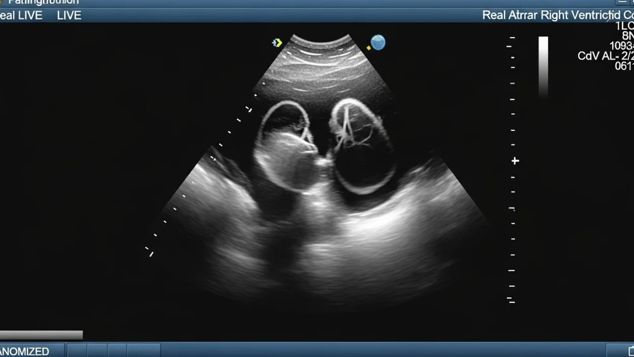
Finally, fetal echocardiograms focus on the heart of an unborn baby. They can detect congenital heart defects before birth, allowing doctors to plan necessary care. Additionally, Doppler and 3D echocardiograms provide enhanced imaging and detailed blood flow analysis, helping physicians assess complex heart conditions more accurately.
By understanding the different types of echocardiograms, patients can better appreciate why their doctor selects a particular test and how it contributes to heart health evaluation.
What an Echocardiogram Can Show
An echocardiogram provides a detailed view of the heart, helping doctors assess both its structure and function. One of the primary things it reveals is the size and thickness of the heart chambers. Abnormal enlargement or thickening can indicate conditions such as high blood pressure, heart failure, or cardiomyopathy. The test also evaluates the motion and strength of the heart muscle, showing how effectively the heart pumps blood.
Another critical feature visible on an echocardiogram is the heart valves. The test can detect valve problems, such as stenosis (narrowing), regurgitation (leakage), or prolapse, which can affect blood flow and overall heart efficiency. Using Doppler imaging, the echocardiogram measures blood flow and pressure within the heart, revealing areas where blood may be moving too quickly, too slowly, or in the wrong direction.
Echocardiograms can also detect fluid around the heart (pericardial effusion) or the presence of blood clots or masses that could cause complications. For children and adults with congenital heart defects, the test identifies structural abnormalities such as septal defects or malformed valves.
In addition, advanced echocardiography techniques, including 3D imaging, allow doctors to observe the heart from multiple angles and gain precise measurements. By providing a comprehensive view of the heart’s chambers, valves, muscles, and blood flow, an echocardiogram is an invaluable tool for diagnosing heart conditions, monitoring ongoing heart health, and guiding treatment decisions.
Common Heart Conditions Detected by an Echocardiogram
Echocardiograms are invaluable in diagnosing a wide range of heart conditions, helping doctors identify issues early and guide treatment plans. One of the most common uses is detecting heart failure, where the heart cannot pump blood efficiently. The test measures ejection fraction, which shows how much blood the left ventricle pumps with each beat, providing critical insight into heart function.
Valve diseases are also frequently diagnosed with echocardiography. These include stenosis (narrowed valves that restrict blood flow), regurgitation (leaky valves allowing blood to flow backward), and prolapse (valve flaps bulging abnormally). Detecting these conditions early can prevent complications such as heart enlargement or arrhythmias.
Cardiomyopathy, a disease of the heart muscle, is another condition an echocardiogram can reveal. The test can show thickened, weakened, or stiffened heart muscles that affect the heart’s ability to pump efficiently.
For individuals with congenital heart defects, echocardiograms identify structural abnormalities present from birth, such as septal defects (holes in heart walls) or malformed valves. The test can also detect blood clots or masses inside the heart that may increase the risk of stroke or other complications.
Other conditions visible through echocardiography include pericardial effusion (fluid around the heart), pulmonary hypertension (high blood pressure in the lungs), and endocarditis (infection of the heart lining or valves). By revealing these conditions, echocardiograms play a critical role in both diagnosis and ongoing monitoring of heart health.
Why Your Doctor May Recommend an Echocardiogram
Doctors recommend an echocardiogram for a variety of reasons, depending on a patient’s symptoms, medical history, and risk factors. One of the primary reasons is to evaluate symptoms of heart disease, such as shortness of breath, chest pain, palpitations, or unexplained fatigue. These symptoms may indicate underlying issues like heart failure, valve problems, or cardiomyopathy, which an echocardiogram can help diagnose.
Patients with a history of heart disease or risk factors—including high blood pressure, diabetes, or a family history of heart problems—may also be advised to undergo echocardiography. Regular monitoring allows doctors to track heart function over time and adjust treatments as needed.
Echocardiograms are commonly used before or after heart surgery to assess the condition of the heart and valves. They can help plan surgical procedures and monitor recovery after interventions. Similarly, doctors use echocardiograms to monitor the effectiveness of medications for heart conditions, such as drugs for heart failure or high blood pressure, by tracking improvements or changes in heart function.
In some cases, an echocardiogram is performed to detect congenital heart defects in children or unborn babies, providing early information that can guide treatment planning. Additionally, stress echocardiograms are used to evaluate how the heart performs under physical activity, helping detect coronary artery disease that may not be visible at rest.
Overall, an echocardiogram is a versatile and non-invasive tool that gives doctors essential insight into the heart’s health, aiding in diagnosis, treatment, and ongoing monitoring.
How to Prepare for an Echocardiogram
Preparing for an echocardiogram is generally straightforward, as it is a non-invasive and safe test. For most standard transthoracic echocardiograms (TTE), no special preparation is required. Patients can eat, drink, and take medications as usual. Wearing comfortable clothing that can be easily removed or adjusted around the chest is recommended, as the technician will need access to the area for the transducer.
For a transesophageal echocardiogram (TEE), preparation is slightly more involved. Because the procedure involves inserting a probe into the esophagus, patients may be asked to fast for several hours beforehand to ensure an empty stomach and reduce the risk of discomfort or nausea. Doctors may also advise stopping certain medications temporarily, particularly blood thinners, but only under medical guidance. Mild sedation is often provided to keep the patient relaxed and comfortable.
Stress echocardiograms require additional preparation. Patients may be instructed to avoid eating or drinking for a few hours before the test and to wear appropriate clothing and shoes for exercise. Certain medications that affect heart rate may need to be paused, but this should always be done under a doctor’s guidance.
It’s also important to inform your doctor about any medical conditions, allergies, or prior heart procedures before the echocardiogram. Bringing a list of current medications can help ensure accurate testing and reduce risks.
By following these preparation steps, patients can ensure the echocardiogram is accurate, safe, and comfortable, allowing doctors to obtain the best possible images and information about heart health.
What Happens During an Echocardiogram
An echocardiogram is a straightforward and generally painless procedure that provides real-time images of the heart. For a transthoracic echocardiogram (TTE), the patient lies on an examination table while a technician applies a special gel to the chest. This gel helps transmit sound waves from the transducer, a handheld device that is gently moved over the chest area to capture images of the heart. The patient may be asked to change positions or hold their breath briefly to obtain clearer pictures. The procedure typically takes 30 to 60 minutes and does not require any recovery time afterward.
For a transesophageal echocardiogram (TEE), the procedure involves inserting a thin probe into the esophagus, which is located close to the heart. Patients usually receive mild sedation to ensure comfort and minimize gagging. The probe captures highly detailed images, particularly useful for assessing heart valves and detecting blood clots. TEE generally takes slightly longer than TTE and may require a short observation period after the procedure until the sedative wears off.
In a stress echocardiogram, the heart is evaluated under physical or pharmacologic stress. Patients may walk or run on a treadmill, or receive medication to increase heart rate, while the echocardiogram monitors how the heart responds to activity.
Throughout the procedure, a cardiologist or technician monitors the images, ensuring all necessary views are captured. Patients may hear the ultrasound waves as a subtle sound, but the test is painless, non-invasive, and safe for nearly everyone.
Interpreting Echocardiogram Results
Once an echocardiogram is completed, a cardiologist analyzes the images to assess the heart’s structure and function. One of the key measurements is the ejection fraction (EF), which indicates how much blood the left ventricle pumps with each heartbeat. A normal EF typically ranges between 55% and 70%, while lower values may suggest heart failure or weakened heart muscles.
The heart chambers are examined for size and thickness. Enlarged or thickened chambers may indicate conditions such as high blood pressure, cardiomyopathy, or valve problems. The motion of the heart walls is also evaluated, as abnormal movement can signal previous heart attacks or muscle weakness.
Heart valves are closely inspected to ensure they open and close properly. Any narrowing (stenosis) or leaking (regurgitation) is noted, along with its severity. Doppler imaging provides additional information about blood flow, helping identify abnormal patterns, high pressures, or backward flow through the valves.
Echocardiograms can also reveal fluid around the heart (pericardial effusion) or blood clots/masses, which may require further investigation or treatment. For congenital defects, the cardiologist identifies abnormal structures such as septal defects or malformed valves.
It’s important to remember that echocardiogram results must be interpreted within the context of the patient’s overall health, symptoms, and other test results. A normal echocardiogram does not guarantee the absence of all heart problems, but it provides critical information that guides diagnosis, treatment decisions, and ongoing monitoring. Patients should always discuss their results with their doctor to understand their heart health fully.
Limitations of an Echocardiogram
While echocardiograms are highly useful for evaluating heart health, they do have some limitations. One of the main challenges is image quality, which can be affected by factors such as obesity, lung disease, or chest wall abnormalities. In these cases, the ultrasound waves may not penetrate effectively, making it harder to get clear pictures of the heart.
Another limitation is that not all heart conditions are detectable with a standard echocardiogram. For example, small coronary artery blockages or early-stage heart disease may not be visible. In such cases, doctors may recommend additional tests like a stress test, CT scan, MRI, or cardiac catheterization to obtain more detailed information.
Certain advanced procedures, such as transesophageal echocardiograms (TEE) or 3D echocardiography, provide more detailed images, but they may not be suitable for every patient due to discomfort, sedation requirements, or medical conditions that limit their use.
Additionally, interpretation of echocardiogram results requires expertise. Small abnormalities may be overlooked or misinterpreted if the images are unclear or the technician lacks experience. Therefore, results are always reviewed by a cardiologist who considers the patient’s symptoms, medical history, and other diagnostic tests.
Despite these limitations, echocardiograms remain a safe, non-invasive, and invaluable tool for assessing heart structure, valve function, and blood flow. Understanding their limitations helps patients set realistic expectations and ensures doctors use complementary tests when necessary for a complete evaluation.
Risks and Safety of an Echocardiogram
Echocardiograms are widely regarded as a safe and non-invasive diagnostic test for evaluating heart health. Most patients experience no discomfort, and there is generally no recovery time required after the procedure.
For standard transthoracic echocardiograms (TTE), the risk of complications is extremely low. Patients may feel minor pressure from the transducer or slight coldness from the gel applied to the chest, but these sensations are temporary and harmless.
Transesophageal echocardiograms (TEE) carry slightly higher risks because they involve inserting a probe into the esophagus. Sedation is often used, so patients may experience temporary drowsiness or a mild sore throat afterward. Rare complications, such as difficulty swallowing or minor irritation of the esophagus, can occur but are uncommon.
Stress echocardiograms, which involve exercise or medication to increase heart rate, also have minimal risks. However, patients with certain heart conditions may require close monitoring to prevent abnormal heart rhythms or chest discomfort during the test.
Overall, echocardiograms are considered very safe, with the benefits of detailed heart assessment far outweighing potential risks. Patients should always discuss any concerns, allergies, or medical conditions with their doctor before the test to ensure proper precautions are taken and the procedure is tailored to their needs.
FAQs:
1. How long does an echocardiogram take?
A standard transthoracic echocardiogram usually takes 30 to 60 minutes, while more advanced procedures like TEE or stress echocardiograms may take slightly longer due to preparation and monitoring.
2. Does an echocardiogram hurt?
No. Most patients feel only mild pressure from the transducer or coldness from the gel. For TEE, mild throat discomfort may occur, but it is temporary.
3. Can an echocardiogram detect a heart attack?
While it cannot detect all heart attacks directly, an echocardiogram can show areas of the heart muscle that are not contracting properly, which may indicate past or recent damage.
4. How often should I get an echocardiogram?
Frequency depends on your heart health, risk factors, and doctor’s recommendations. Patients with chronic heart conditions may need periodic monitoring, while others may only need one test if symptoms arise.
5. Are there any risks?
Risks are minimal. Standard echocardiograms are virtually risk-free. TEE carries a slight risk of throat irritation or discomfort from sedation, and stress tests require careful monitoring for patients with heart disease.
6. Can echocardiograms replace other heart tests?
No. While they provide detailed structural and functional information, echocardiograms are often used alongside ECGs, stress tests, or imaging studies for a complete evaluation.
Conclusion
An echocardiogram is a safe, non invasive, and highly effective tool for assessing heart health.
It provides detailed information about the heart’s structure, valves, chambers, and blood flow, helping doctors detect conditions such as heart failure, valve diseases, cardiomyopathy, and congenital defects.
With different types of echocardiograms available transthoracic, transesophageal, stress, and fetal physicians can choose the best method to gather accurate information for each patient’s needs.
While the test is generally risk-free, it has some limitations, including image quality variations and the inability to detect certain heart conditions without complementary tests.
Proper preparation and understanding of the procedure can make the experience comfortable and efficient.
By interpreting echocardiogram results in the context of overall health, doctors can make informed decisions about treatment, monitoring, and preventive care.
Ultimately, echocardiograms play a crucial role in maintaining heart health and supporting early diagnosis and effective management of heart conditions.

I’m the mind behind Jokesattack.com, your go-to place for daily laughs, clever puns, and hilarious memes. Spreading smiles one joke at a time!